Research Areas
- EEG-fMRI fusion of Steady-State or Event-Related Brain Potential
- Brain connectivity
- Multilinear Methods
- Localization of epileptic foci using source reconstructed EEG data
- Source localization of spatio-temporally decomposed topographic EEG maps
Projects
1. EEG-fMRI fusion with Multilinear Methods
 Project Coordinator: Prof. Dr. Ahmet Ademoğlu,
Researcher: Esin Karahan
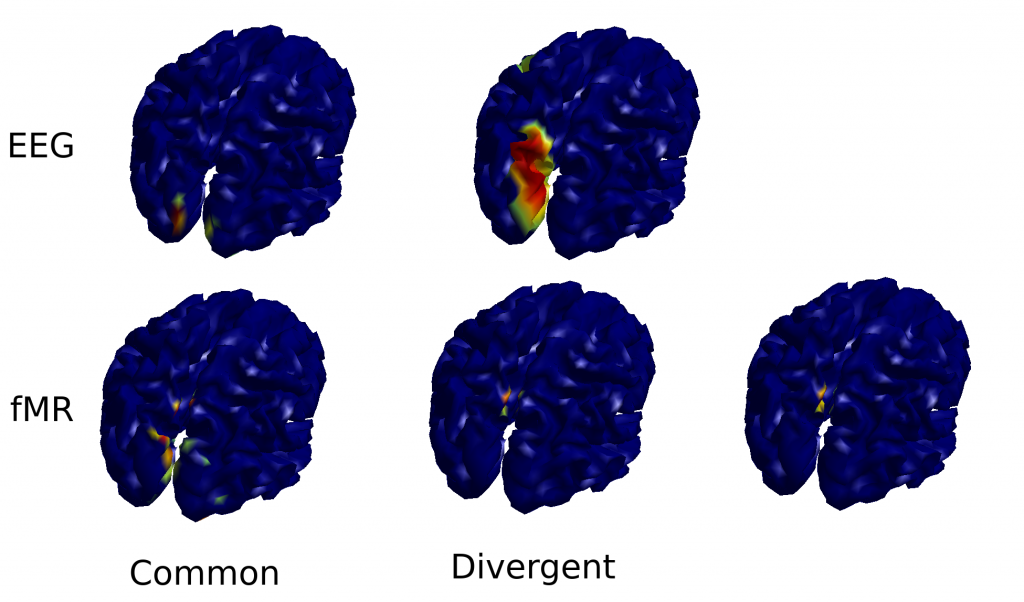
Project Summary: Scalp EEG signals are direct measure of brain electric activity by reflecting the postsynaptic cortical currents generated by the large pyramidal neurons which are located perpendicularly to the cortical surface. Functional MR imaging (fMRI) is the other way of measuring brain functions reflecting the oxygen metabolism of the brain. Temporal resolution of BOLD (blood-level-oxygen-dependent) signal measured with fMRI is confined to the time course of slow evolving hemodynamic activity (~ 10 s) while exhibiting high spatial resolution in terms of millimeters. Despite its low spatial resolution, scalp EEG measures brain function with a temporal resolution in terms of miliseconds. By compromising different imaging modalities, fusion of EEG and fMRI on a common space and time scale is one of the current problem of neuroimaging to reveal the complex dynamics of brain function and neuronal interactions. In this study, a novel EEG-fMRI fusion approach based on multilinear methods will be developed and applied to clinical and neurophysiological data. In this way, different platforms representing brain activity data will be integrated on the same spatial and temporal scales. Proposed approach will contribute to studies on brain functions and representation of memory, attention, information processing mechanisms in the brain. Proposed approach has a wide aplication area from diagnosis of neurological diseases to brain-computer interface studies.
Keywords: EEG-fMRI fusion, multilinear methods, parallel factor analysis, inverse problem, penalized regression
Funded by Bogazici University Scientific Research Project Council (2012 – 2014)
2. Localization of Epileptic Sources in Surgical Planning by using Deep EEG Electrodes (Derin EEG Elektrotlarının Beyin Dokusuna Yerleştirimine Dayalı Cerrahi Planlamada Epileptik Bölgelerin Kaynak Görüntülemesi)
Project Coordinator: Prof. Dr. Ahmet Ademoğlu
Researcher: A. Deniz Duru
3. Investigation of the Neuro-vascular Coupling in the Brain: The Study and Modeling of the BOLD Counterparts of Steady-State Evoked Potentials using EEG-fMRI Technique
Project Coordinator: Prof. Dr. Tamer Demiralp, Istanbul Medical School, Physiology Department
Project Summary: Joint use of non-invasive measurement techniqeus of brain function, the electroencephalogram (EEG) with high temporal resolution and the functional magnetic resonance imaging (fMRI) with high spatial resolution, provides an important possibility for understanding both structural and dynamical properties of neural processes underlying human brain function. Complementary use of these techniques requires a correct model of the electro-vascular coupling between the EEG activities and the vascular response. Present models employed in the EEG-fMRI integration studies assume a relationship between the level of the neuronal activity and the oxygen transport to the tissue. However, changes in the EEG signal do not reflect directly the level of neural activity but synchrony. In this project, the correlations between the synchronized EEG patterns and the fMRI have been investigated in the visual modality. Steady-state visual evoked potentials (SSVEP) were used to obtain synchronized EEG patterns that remain constant along the slower simultaneous fMRI response, and correlations between changes of the EEG and fMRI due to the stimulation frequency were investigated. Although a strong correlation exists between the EEG and fMRI changes for stimulation frequencies in the beta frequency range, the correlation disappears when stimuli were applied with frequencies corresponding to the alpha and gamma frequency ranges of the EEG. This specific difference among the frequency bands of stimuli has been explained by the differences of the synchronization mechanisms and required excitatory and inhibitory inputs to synchronize oscillatory activities in the visual cortex in different frequency ranges. In EEG/fMRI integration studies it should be taken into account that both signals do not reflect same neural processes, but as long as experimental variables are used that affect both signals, and symmetrical analysis approaches are employed instead of asymmetrical approaches such as defining the solution space of one modality using information obtained from the other, the presence as well as the absence of a correlation between them can shed light on the underlying neural processes.
Keywords: fMRI, Steady-state Evoked Potentials, EEG, Neuronal synchrony, BOLD


